1MD, PhD I.R.C.C.S. Giovanni Paolo II, Bari, Italia
2Consulente Tecnico di Istituto Nazionale Biosistemi e Biostrutture Bologna Italy
Keywords: cellular vibrational coherence, early tumor transformation, biophysical biomarkers, Kuramoto model, Raman spectroscopy.
Abstract: The cell can be conceived as a complex dynamic system composed of interconnected oscillatory domains, in which biochemical, mechanical, and energetic processes generate coordinated vibrational patterns. Under physiological conditions, the synchronization among these domains—encompassing the cytoskeleton, membrane, intracellular compartments, and metabolic dynamics—contributes to structural stability, metabolic efficiency, and proper signal transduction. In this context, cellular vibrational coherence can be interpreted as an emergent indicator of the physiological state of the biological system. Tumor transformation introduces profound alterations in cellular organization: cytoskeletal remodeling, changes in mechanical stiffness, nuclear reorganization, and metabolic reprogramming associated with the Warburg effect modify the dynamic equilibrium of the cell (Hanahan & Weinberg, 2011). These changes may lead to a progressive loss of synchronization among intracellular oscillatory domains, generating a vibrational signature distinct from that of healthy cells.
In this work, we propose the evaluation of cellular vibrational coherence as a potential biophysical biomarker of pathological transition. The theoretical model considers the cell as a network of coupled oscillators, describable through principles of nonlinear dynamics and phase synchronization, similarly to what is formalized in the Kuramoto model (Kuramoto, 1984; Strogatz, 2003). In this context, a quantitative index of cellular vibrational coherence (CVC) is proposed, capable of expressing the degree of synchronization among the various intracellular oscillatory domains
The approach integrates advanced imaging and vibrational spectroscopy techniques—such as Raman spectroscopy and Brillouin microscopy—with computational biophysical models, enabling the non-invasive mapping of cellular oscillatory dynamics (Puppels et al., 1998; Scarcelli & Yun, 2010). The loss of vibrational coherence can be interpreted as a dynamic transition of the cellular system, opening new perspectives for predictive biomedicine, the development of innovative biophysical biomarkers, and the implementation of high-sensitivity diagnostic technologies for preventive oncology and therapeutic monitoring.
Early Cancer Diagnosis
Early cancer diagnosis represents one of the major challenges of contemporary medicine. Current diagnostic approaches are primarily based on the identification of genetic alterations, molecular biomarkers, or morphological changes in cells. However, such modifications often become detectable only when the tumor transformation process is already advanced.
In recent years, the need has emerged to identify earlier indicators that reflect systemic changes preceding evident phenotypic alterations. From this perspective, the cell can be interpreted not only as a biochemical system, but as a complex dynamic system characterized by nonlinear interactions among structural, metabolic, and mechanical components. This view is situated within the context of systems biology, according to which the emergent properties of biological systems arise from the dynamic interaction of their components (Kitano, 2002). Similarly, principles of complex systems and self-organization suggest that the physiological state of a living system may be associated with specific patterns of dynamic order.
In this context, we propose that the state of cellular health is associated with a condition of vibrational coherence, whereas tumor transformation would correspond to a progressive loss of synchronization among intracellular oscillatory domains.
The Cell as a System of Coupled Oscillators
Many cellular processes exhibit oscillatory or periodic characteristics, including cytoskeletal dynamics, metabolic fluctuations, mitochondrial activity, and movements of the plasma membrane. These phenomena suggest that the cell can be modeled as a network of interconnected biological oscillators.
Within the framework of nonlinear dynamics, the collective behavior of systems of coupled oscillators can be described using mathematical models of synchronization. Among these, one of the most well-known is the Kuramoto model, developed to describe the transition between coherent and incoherent states in complex systems composed of many oscillators with different natural frequencies (Kuramoto, 1984; Strogatz, 2003). According to this model, when the coupling among oscillators exceeds a critical threshold, the system spontaneously transitions from a disordered state to a synchronized one. This phenomenon is observable in natural systems, including neuronal circuits and oscillating biochemical systems.
Applying this approach to the cell, each oscillatory domain can be characterized by a dynamic phase θᵢ. The overall degree of synchronization of the systemcan be expressed through an order parameter:
Here: N is the number of oscillatory domains considered, θⱼ is the phase of the j-th oscillator, i is the imaginary unit, and |⋅| denotes the modulus of the resulting complex number.modulus of the resulting complex number.
The resulting parameter, called Cellular Vibrational Coherence (CVC), ranges from 0 to 1:
- CVC values close to 1 indicate high synchronization (stable physiological state)
- CVC values close to 0 indicate a loss of dynamic coherence (disordered state)
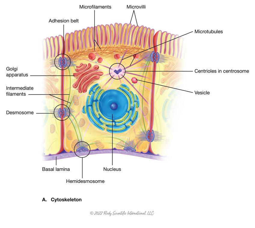
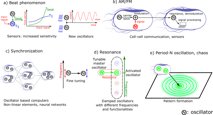
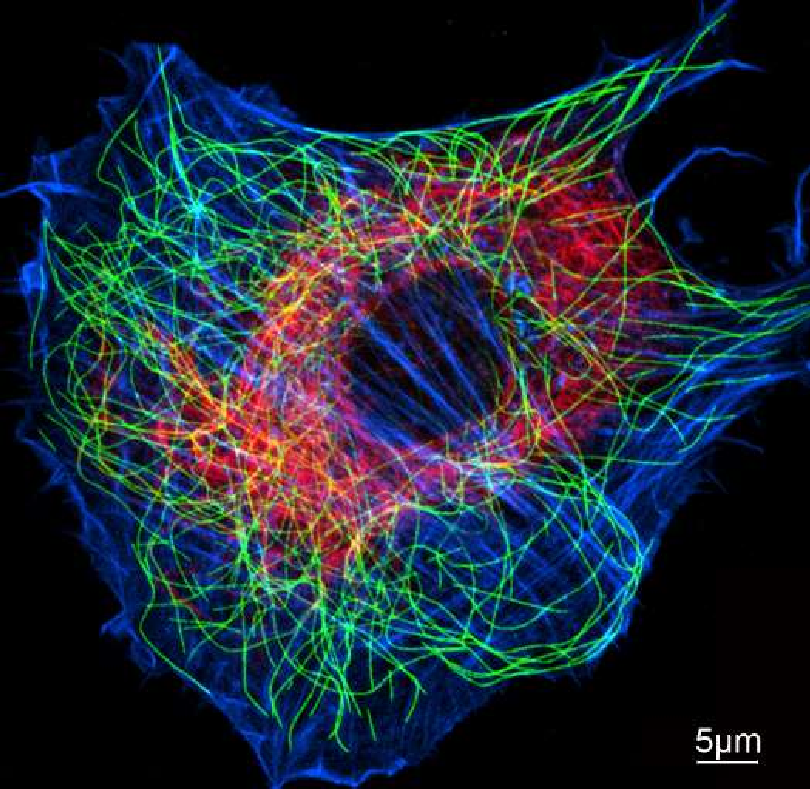
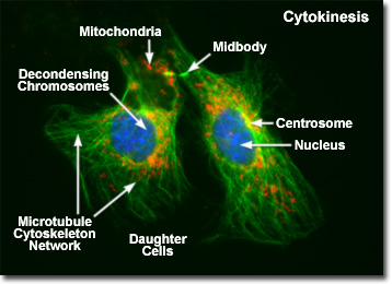
Figure 1 – Conceptual Model of Cellular Vibrational Coherence
In the physiological state, the various oscillatory domains of the cell—including the cytoskeleton, plasma membrane, intracellular compartments, and metabolic activity—exhibit high dynamic synchronization, corresponding to elevated values of the Cellular Vibrational Coherence (CVC) parameter.
During tumor transformation, structural and metabolic changes—including cytoskeletal remodeling, alterations in cellular stiffness, and metabolic reprogramming—can cause a progressive loss of synchronization among these oscillatory domains, leading to a decrease in the CVC parameter and the emergence of a distinctive vibrational signature.”
Loss of Vibrational Coherence in Tumor Transformation
Tumor transformation is associated with numerous structural and functional alterations of the cell:
- Cytoskeletal remodeling
- Changes in cellular stiffness
- Alterations of chromatin and nuclear organization
- Metabolic reprogramming (Warburg effect) (Hanahan & Weinberg, 2011)
These modifications affect the mechanical and dynamic properties of the cell, leading to a progressive loss of synchronization among intracellular oscillatory domains (Fleury et al., 2020). The result is an altered vibrational signature, which can be detected using imaging and spectroscopy techniques.
Technologies for Measuring Cellular Vibrational Dynamics
Among the technologies potentially applicable to the study of cellular vibrational coherence:
- Raman spectroscopy, which detects specific molecular vibrations (Puppels et al., 1998)
- Brillouin microscopy, which measures the mechanical and viscoelastic properties of tissues (Scarcelli & Yun, 2010)
- High-resolution imaging of intracellular fluctuations
Integration with computational models can enable the reconstruction of the cell’s dynamic spectrum and the estimation of the CVC parameter
Implications for Early Diagnostics
The loss of vibrational coherence in cells may occur before traditional morphological alterations, suggesting that the CVC parameter could serve as an early biomarker. This opens promising perspectives: it could be used for non-invasive cancer screening, to identify early-stage unstable cellular states, and to monitor the effectiveness of therapeutic interventions.
In this context, the analysis of vibrational coherence complements the philosophy of predictive and personalized medicine, providing information that integrates with traditional molecular biomarkers and contributing to a more comprehensive assessment of cellular functional status.
Experimental Perspectives
The CVC model allows for concrete predictions: tumor cells should exhibit a significantly lower CVC compared to healthy cells, and pharmacological modulation of cytoskeletal or metabolic dynamics could temporarily restore vibrational coherence. These hypotheses are testable using high-resolution imaging and spectroscopy techniques, paving the way for experimental validation of the proposed paradigm.
Conclusions
The cell can be interpreted as a dynamic system of coupled biological oscillators, whose synchronization reflects the physiological state. Tumor transformation can be described as a dynamic transition from a coherent to a disordered state. Measurement of cellular vibrational coherence represents a new frontier in cancer research, with potential applications in the development of innovative biophysical biomarkers and high-sensitivity diagnostic technologies
Bibliography
Fleury, J. B., et al. (2020). Cellular mechanical properties and cancer: The role of cytoskeleton and nucleus in cell deformation and migration. Biophysical Journal, 118(2).
Hameroff, S. R., & Penrose, R. (2014). Consciousness in the universe: A review of the “Orch OR” theory. Physics of Life Reviews, 11(1), 39–78.
Hanahan, D., & Weinberg, R. A. (2011). Hallmarks of cancer: The next generation. Cell, 144(5), 646–674.
Kitano, H. (2002). Systems biology: A brief overview. Science, 295(5560), 1662–1664.
Kuramoto, Y. (1984). Chemical oscillations, waves, and turbulence. Springer.
Puppels, G. J., et al. (1998). Raman spectroscopy in cancer diagnostics. Nature Biotechnology, 16(2), 168–172.
Scarcelli, G., & Yun, S. H. (2010). Brillouin microscopy: Non-contact mechanical imaging. Nature Photonics, 2, 39–43.
Strogatz, S. H. (2003). Sync: How order emerges from chaos in the universe, nature, and daily life. Hyperion.